Recognition of ancient shoreface deposits. Facies, facies sucessions, and associations. An example from the cretaceous gallup clasticwedge, New México
Published 1988-11-09
How to Cite
Abstract
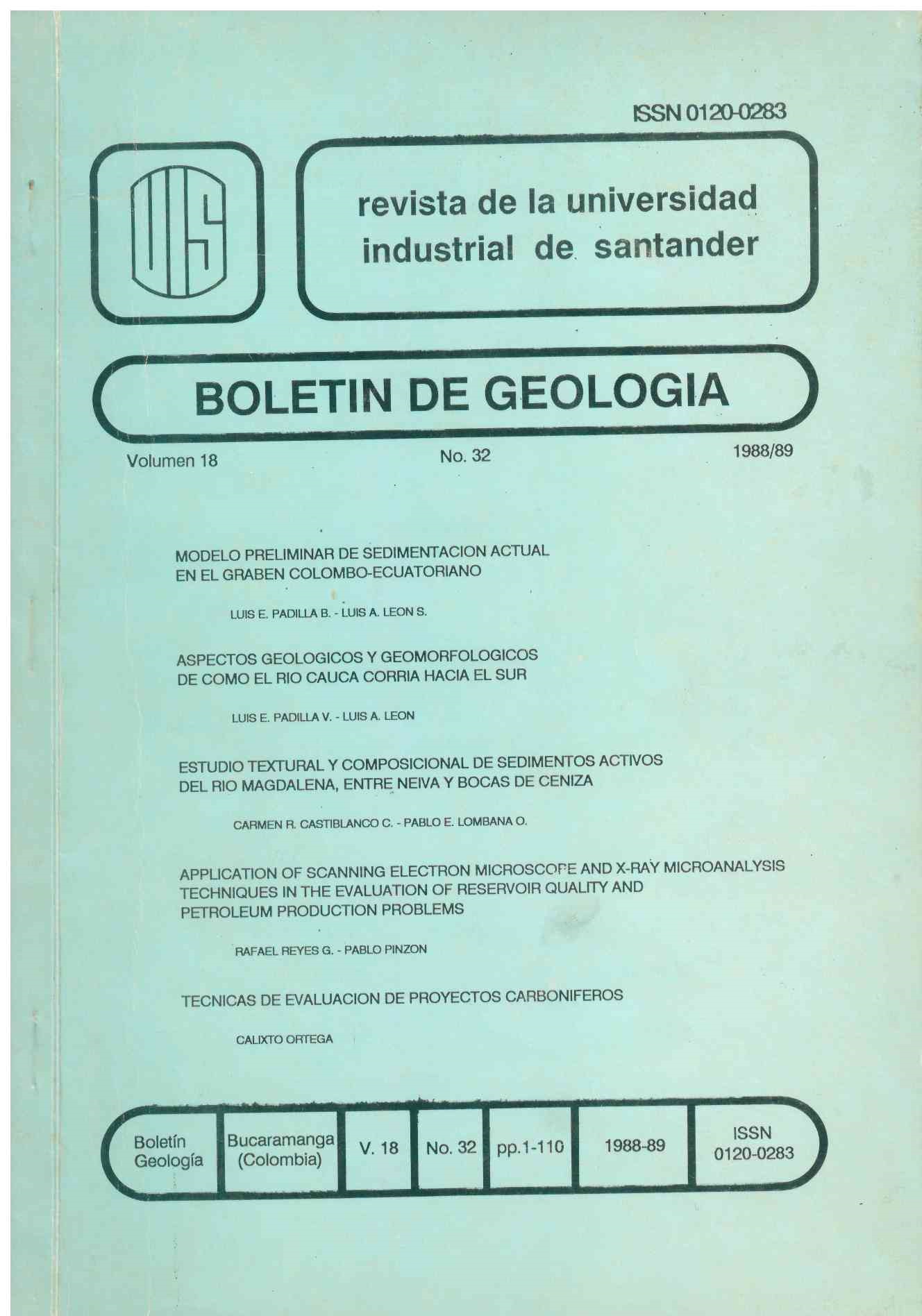
this report summarises the findings of an introductory program to test the application of the scanning electro microscope (SEM) energy dispersive X-ray microanalysis system (EDX) for geological investigation of reservoir quality and petroleum production problems. The three SEM techniques used were.
1.Secondary-electron imaging (SEI), 2. backscatter-electron imaging (BEI), and 3. cathodoluminescence (CL)
Analysis by SEI provides three dimensional images of pore and grain surfaces, from which the following may be defined:
-Pore geometry, -the distribution of pore-throat blocking phases, -phases capable of damaging the reservoir, and -evidence for paramagnetic timing.
BEI analysis by SEM provides detail of compositional differences and is therefore principally of use as a tool for assessment of diagenetic problems. This techniques is also of use in examination of pore system and provides a means of direct comparison wich optional microscopy studies.
CL-imaging by SEM is as yet an unproven technique, but has potential for analysis of carbonates and possibly quartz cements. The SEM, when used in conjunction with standard thin section microscopy, X-ray difraction analysis, core analysis and sedimentological data provides a highly sophisticated but readily masterable tool for interpreting the geological controls on reservoir properties and predicting the lateral variation the may be expected in a reservoir.
